Equipment
IncuCyte® S3 Live-Cell Imaging and Analysis System
Incucyte® S3 Live-Cell Analysis System for automatic long-term acquisition and analysis of cells in a physiologically relevant environment.
-
Two-channel fluorescence (green and red) as well as phase contrast imaging
-
4X, 10X, 20X objectives on an automated turret
-
Support for three interchangeable vessel trays compatible with flask, dishes, slides and microplates - up to six microplates in parallel. Supports multiple users with different protocols at the same time
-
Multi-user, multi-application support through remote network capability
-
Robust application suite
Long term monitoring of cell health to complex functional assays utilizing of up to two totalfluorescence and phase imaging channels simultaneously.
NIKON A1R CONFOCAL TIRF STORM MICROSCOPE (MOORES CANCER CENTER ROOM 5365)
Super Resolution Imaging
Using the Nikon A1R STORM feature, resolution beyond the normal diffraction limit of light can be achieved by stochastic optical reconstruction. Multiple images are acquired during rapid sequential excitation/deactivation of fluorescent probes. Only a small percentage of the fluorescent molecules are switched on during any one activation cycle allowing individual molecules to be clearly identified in each frame. Since images are reconstructed from signals generated from single fluorophores, this allows precise localization of individual molecules in contrast to standard microscopy in which the image is the average of all fluorophore signals. This method produces images with resolution down to 20 nm in the x-y plane, and 50 nm in the z-axis – significantly greater than regular widefield or confocal microscopy.
Total Internal Reflection Fluorescence (TIRF) microscopy
In TIRF microscopy an evanescent wave from light reflected at a critical angle selectively illuminates a field of view a few hundred nanometers at the cell-glass coverslip interface. This results in a high signal to noise ratio that is optimal for studying dynamic cell-surface events.
Laser Scanning Confocal Microscopy
Optical sectioning is achieved by passing light through a pinhole that generates an image that is devoid of out of focus light typically found in wide field microscopy.
Spinning Disk Confocal Microscopy
The spinning disk uses an array of pinholes on a disk spinning at high speed to create a virtual pinhole. Images are acquired on a sCMOS camera giving an instantaneous full frame image as in widefield microscopy. This results in acquisition at relatively higher speeds as compared to the laser scanning mode making it ideal for live cell and fast confocal imaging.
- Nikon Ti microscope with integrated autofocus (Perfect focus) that maintains focus when using glass slides, plastic culture dishes, or multiwell plates
- Automated XY and Z stage (including a fast piezo Z)
- Andor iXon+DU897, EMCCD (512x512 pixels with readout of 30 full frames per second) for low light applications such as TIRF, FRAP, and single molecule imaging applications such as STORM and PALM
- Yokogawa CSU10 spinning disk scanner unit with attached Andor high speed sCMOS camera (Zyla 5.5 megapixel with readout of 100 full frames per second) for applications requiring a higher resolution and speed such as live cell confocal imaging.
- A1R hybrid confocal scanner incorporates two independent galvo scanning systems, a high-resolution (4096x4096) scanner, and a high speed resonant scanner (512x512) for rapid confocal scanning of live cells during simultaneous photobleaching or photoactivation for photokinetic studies
- LU4 four-laser AOTF unit with 405, 488, 561, and 647 lasers
- Filters for DAPI, CFP, FITC/GFP, YFP, Rhodamine/Texas red/mCherry, Cy5 and Cy7. Additional narrow bandpass filters for Alexa 488, Alexa 523, Alexa 555, Alexa 594, Alexa 647 and Alexa 750. Using a combination of these narrow bandpass filters and the Huygens spillover correction software, it is possible to image 6 channels plus DAPI on the same slide with the 4 lasers.
- Lumencore solid state light source for wide field illumination and fast switching
- Plan Apo 10, 20 and 40X dry objectives, 40X oil and 60 and 100X high NA (1.49NA) TIRF objectives
- TokaiHit stagetop incubator with heating and carbon dioxide incubation for live cell work
- The microscope is located in a BSL2 facility
- Super resolution microscopy using stochastic optical reconstruction methods such as N-STORM, D-STORM, or PALM (Photo Activation Localization Microscopy)
- Three-color 2D & 3D-STORM with conventional dyes such as Alexa488, Alexa568, and Alexa647
- 3D super resolution imaging that allows accurate localization of sub-cellular components
- TIRF microscopy of dynamic cell-surface events, such as endocytosis, exocytosis, focal adhesion dynamics, integrin and receptor mediated signaling, invadopodial/podosome dynamics
- Laser scanning confocal microscopy of fixed and live samples
- Photokinetics, - simultaneous photo activation or photo bleaching (FRAP) and imaging
- Forster Resonance Energy Transfer (FRET)
- Generation of high quality 3D and multi color montages with the automated XYZ stage
- High speed, or low light level imaging
- Multi dimensional fast time-lapse imaging
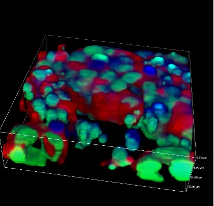
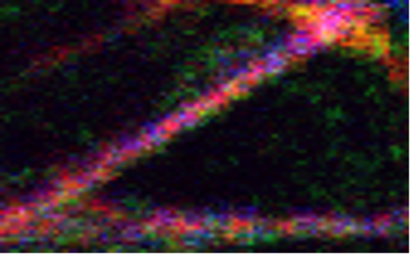
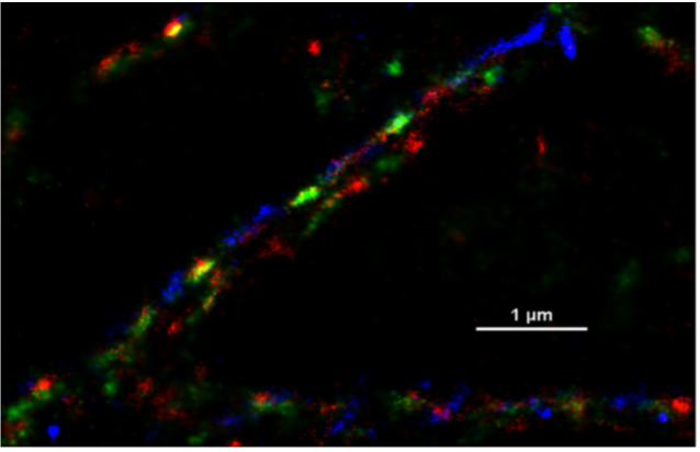
Arrangement of Intermediate Filament Proteins in Glioblastoma Cells. Individual filaments, and the arrangement of 3 intermediate filament proteins therein are clearly resolved by 3 color STORM. Vimentin (green) and Nestin (red), GFAP (blue).
INFORMATIONAL VIDEOS ABOUT STORM AND PALM
This YouTube video describes the principles of STORM:
These YouTube videos provide additional information:
IVIS 200 IN VIVO BIOLUMINESCENCE / FLUORESCENCE IMAGING (MOORES CANCER CENTER VIVARIUM)
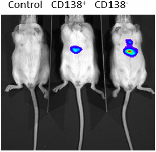
The XENOGEN IVIS 200 Imaging System can be used to image both bioluminescence and fluorescence non-invasively in living animals, and to perform quantitative in vitro and in vivo assays using reporter cells tagged with a wide range of bioluminescent or fluorescent probes. The system uses a novel Xenogen technology in vivo biophotonic imaging to allow researchers to use real-time imaging to monitor and record cellular and genetic activity within a living organism.
- Integrated fluorescence system (400–900 nm) allows easy switching between fluorescent and bioluminescent spectral imaging applications
- Laser scanner provides 3D surface topography for single-view diffuse tomographic reconstructions of internal sources
- Spectral imaging uses measurement data from a sequence of images filtered at different wavelengths, ranging from 560 nm to 660 nm, to determine the depth and location of a bioluminescent reporter
- Excitation and emission filters for GFP, DsRed, Cy 5.5, and ICG in addition to a set of four background filters for subtraction of tissue autofluorescence
- A 26 mm square back-thinned 16 bit 2048x2048 pixel CCD camera cryogenically cooled to –105° C minimizes electronic background, and maximizes sensitivity.
- Non-invasive imaging of distribution of cells tagged with bioluminescent or fluorescent markers following injection in live animals over time
- Localization of tumor or non-tumor cells in 3D in live animals, e.g. invasion and metastasis of tumor cells
- Quantitative comparison of fluorescence or bioluminescence for tracking treatment efficacy over time
SAMPLE IMAGES
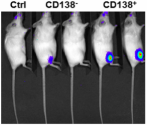
Each mouse was transplanted intrafemorally to the right femur with 53,00 sorted cells isolated from fresh bone marrow biopsy of a multiple myeloma patient and transduced with GLF containing lentivirus. Bioluminescence signals were detected as early as 4 weeks post-transplantation.
Live IVIS image was taken 4 weeks after intrahepatical transplantation of H929 cells transduced with GLF lentivirus into neonates.
Bioluminescent Monitoring of Microenvironmental Effects on Multiple Myeloma Engraftment in a Human Xenograft Mouse Model using IVIS200. Images provided courtesy of Christina C.N. Wu, and Dr. Dennis Carson
KEYENCE BZ-X700 ALL IN ONE FLUORESCENCE MICROSCOPE (MOORES CANCER CENTER ROOM 5317)
Fluorescence generated by excitation of fluorophores is captured by a CCD camera
- No darkroom required
- Fully motorized control including large motorized stage that supports multi-well plates
- High-resolution 2.8 megapixel monochrome/color CCD camera
- Narrow band-pass filters to image six fluorophores (DAPI, FITC, TRITC, Cy5 and Cy7 or IR dye800
- Objective lenses include 2x, 4x, 10x, 20x, and 40X dry objectives, 60X and 100x oil objectives
- Hybrid cell count/Macro cell count
- Imaging both, fluorescence staining, as well as slides stained with chromogenic dyes
- Motorized stage allows capture of fully-focused XYZ stitched images
- Multidimensional capture feature allows automatic capture of multiple wells/samples
- Highly automated, user friendly and simple data analysis (e.g. intensity measurements, counting, etc.) platform allows easy quantitative measurements and recording.
NIKON UPRIGHT FLUORESCENCE MICROSCOPE (MOORES CANCER CENTER ROOM 2304)
Fluorescence generated by excitation of fluorophores is captured by a CCD camera
- Nikon E600 upright fluorescence microscope
- 150W Xenon arc lamp
- narrow band-pass filters to image six fluorophores (DAPI, FITC, Cy3, Red, Cy5 and Cy7) in the same sample
- Objective lenses include 4x, 10x, 20x, and 40X DIC objectives in addition to 60X and 100x oil objectives DIC optics
- 14 bit SPOT Explorer mono/color dual camera that is more sensitive in the infrared region
- Imaging both, fluorescence staining, as well as slides stained with chromogenic dyes
- Imaging tissue samples stained with fluorophores with excitation and emission in the infra red region - an important feature when trying to correlate the observations made in live animals (using the IVIS whole animal imaging system) with events that are occurring at the tissue and cellular level. The existing filters are useful for imaging dyes such as Cy5, Cy5.5, Cy7, Alexa750
SPECTRA OF DYES AND FILTERS
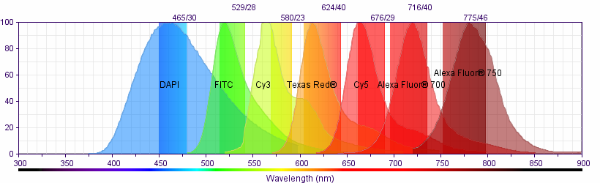
Emission Spectra of recommended dyes and Bandpasses of the emission filters on the Nikon Upright microscope.
NIKON TEACHING MICROSCOPE (MOORES CANCER CENTER ROOM 2304)
Transmitted light illumination of samples stained with chromogenic dyes
- Nikon E600 upright fluorescence microscope
- Spot QE color camera
- Multiple eyepiece ports for teaching or group
- Objectives lenses include 2x, 4x, 10x, 20x and 40x dry, and 60x and 100x oil
- Imaging Histology specimens stained with chromogenic dyes
- Teaching and group observations
Software
-
NIS-Elements 6D imaging for multi-dimensional acquisition and analysis
-
Ai-Denoise and Clarify modules, 2D and 3D Deconvolution and Tracking
-
General Analysis (GA) and GA3 for 2D and 3D image segmentation, counting and measurement. Batch processing in both GA and GA3 modes
-
2D and 3D STORM analysis module with automatic drift correction
-
Photokinetics and FRET modules
-
3D rendering and measurements
-
Compatible with images generated with third party software like QPath, Image scope, etc.
-
Crosstalk and Autofluorescence corrector performs automated estimation and correction of channel bleed-through and autofluorescence correction within images of up to 32 different channels.
-
Estimated crosstalk correction values of all channels are plotted in a crosstalk coefficient matrix which can be further edited and saved to apply to multiple images in batch settings
-
Multiple extractions and segmentation features for brightfield, fluorescence and color (chromogenic) images
-
Range of measurements and segmentation options
-
Batch processing of data
- Cross-platform, user-friendly open-source software for digital pathology and image analysis
- powerful set of tools for working with whole slide images
- U_Quantify is a MATLAB/Python software suite designed by the Danuser lab for live cell image analysis (https://www.danuserlab-utsw.org/)
- Includes a collection of tools for cross correlation between cellular movement and spatio-temporal analysis of signaling, actin polymerization, FRET analysis, 2D and 3D tracking, segmentation, 3D cell surface topology and analysis tools and more.
Custom Software Design
In addition to the above commercial software packages, the facility is greatly enhanced by collaborations with members of the San Diego Supercomputer Center (SDSC). These collaborations have truly changed the way we visualize and analyze 3-dimensional microscopy data sets. Using SDSC software and expertise, users of the Cancer Center's imaging facility have been afforded unique opportunities created by these remarkable groups of computer scientists.